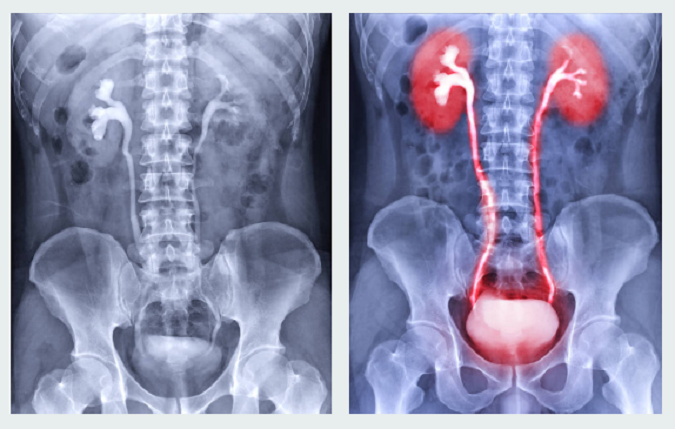
Black: high density of contrast medium. White: low density of
By A Mystery Man Writer
Last updated 15 Jul 2024

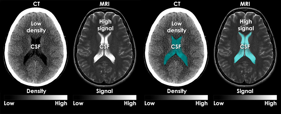
MRI interpretation - What are MRI images?
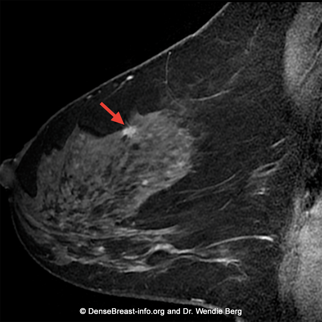
Breast MRI DenseBreast-info, Inc.

Limy bile, Radiology Reference Article

Methodology of the three different aortic measurements in a patient

Chun-Chao Huang's research works Mackay Medical College and other places

Hounsfield Scale - an overview
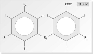
Contrast media

PDF] USE OF CONTRAST MAMMOGRAPHY IN THE DIAGNOSIS OF BREAST CANCER

After Stanford A dissection, the true lumen was compressed by a huge

Phase-contrast virtual chest radiography

After Stanford A dissection, the true lumen was compressed by a huge

Radiographic contrast, Radiology Reference Article
Recommended for you
-
 News - 5 Points to Learn About Contrast Media15 Jul 2024
 -
 To Use Contrast, Or Not Use Contrast: That Is The Question15 Jul 2024
 -
 Radiological Contrast Agents and Radiopharmaceuticals - ScienceDirect15 Jul 2024
 -
 Best Medium for thining GW Washes - + GENERAL PCA QUESTIONS + - The Bolter and Chainsword15 Jul 2024
 -
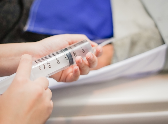
contrast medium injection - Stock Image - C015/2246 - Science Photo Library15 Jul 2024
-
 Ethiodized poppyseed oil-based contrast medium is superior to water-based contrast medium during hysterosalpingography regarding image quality improvement and fertility enhancement: A multicentric, randomized and controlled trial - eClinicalMedicine15 Jul 2024
 -
 Nonionic Contrast Medium - an overview15 Jul 2024
 -
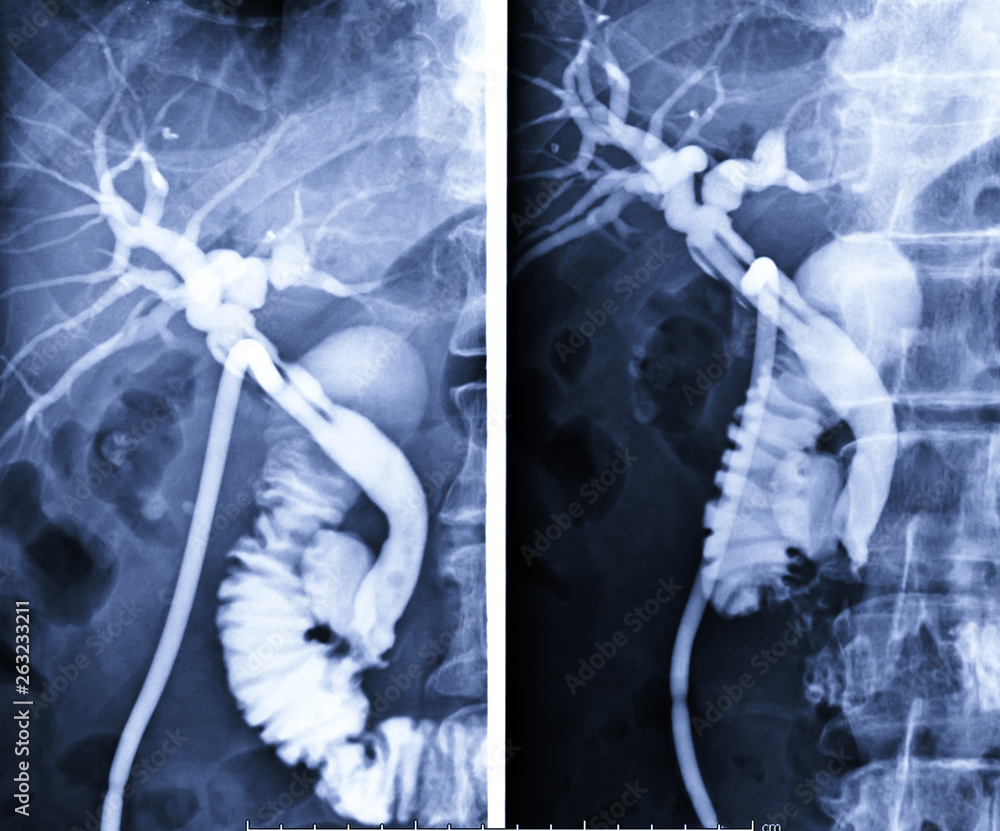 A T-tube cholangiogram is a fluoroscopic procedure in which contrast medium is injected through a T-tube into the patient's biliary tree comparison AP and Oblique view. Stock Photo15 Jul 2024
 -
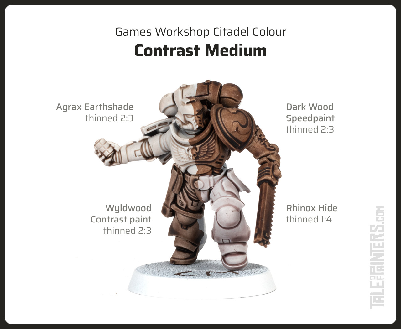
 Citadel Paint: Technical - Contrast Medium15 Jul 2024
 -
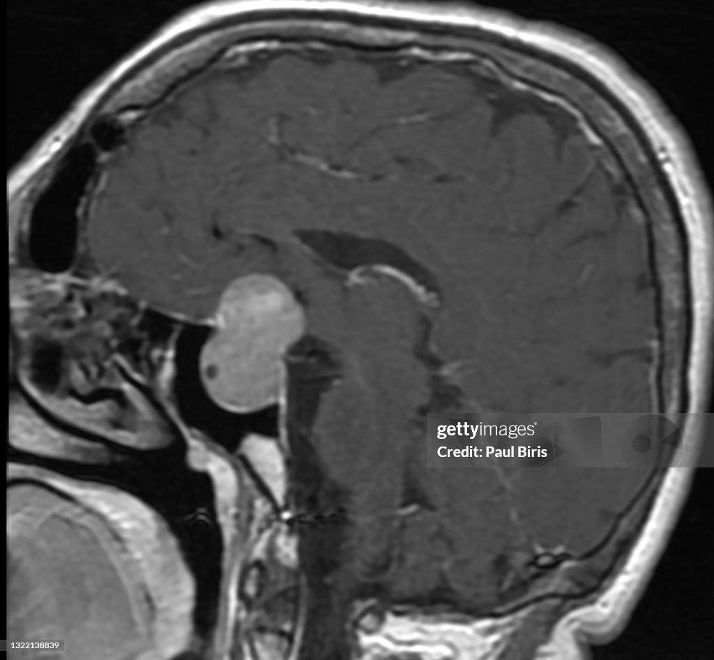 Magnetic Resonance Imaging Contrast Medium Injections Sagittal15 Jul 2024

You may also like
-
 XIFEI Cigar Vcutter Punch Stand 3IN115 Jul 2024
 -
 DragonHawk Extreme V2S Rotary Tattoo Machine Power Supply Traditional Tattoo Needles Grips with Clip Cord Foot Pedal TZ35015 Jul 2024
 -
 Unwrapped Crayons in Bulk - Premium Paperless Crayons with No Paper Wrapper in 9 Colors | Safety Tested Compliant with ASTM D-4236!15 Jul 2024
 -
 Downloads, PE535, United States15 Jul 2024
 -
 Crafting Meat Butcher Paper Disposable Butcher Paper Sheets - Temu15 Jul 2024
 -
 SGHUO Iron on Vinyl Heat Transfer Vinyl 22pcs Includes 16pcs Assorted Colors Sheets and 6pcs Glitter Sheets for T-shirts Works15 Jul 2024
 -
 3 Skeins Yarn Bee Chunky Spiral Almond Bark color Super Jumbo- 715 Jul 2024
 -
 Soarer Black Craft Feathers Bulk - 300pcs 3-5inch Natural Feathers for Wedding Home,Dream Catcher Supplies,DIY Crafts and Halloween Holiday Party(Black)15 Jul 2024
 -
 Janome 7 mm Low Shank Convertible Free Motion Quilting Foot Set - 20200200415 Jul 2024
 -
 Young Living Oregano Essential Oil 15 ml, new sealed15 Jul 2024