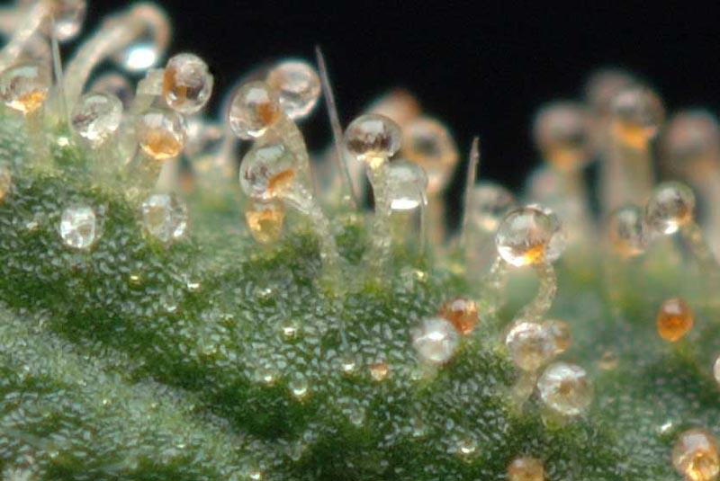
Light microscope (a – c and e) and μ -SXRF (d and f) images of leaf
By A Mystery Man Writer
Last updated 16 Jul 2024


Sascha TAFELSKI, Medical Doctor, PD Dr. med., Charité Universitätsmedizin Berlin, Berlin, Charité, Department of Anesthesiology and Operative Intensive Care Medicine

PDF) Retrograde Lymphatic Spread of Esophageal Cancer: A Case Report
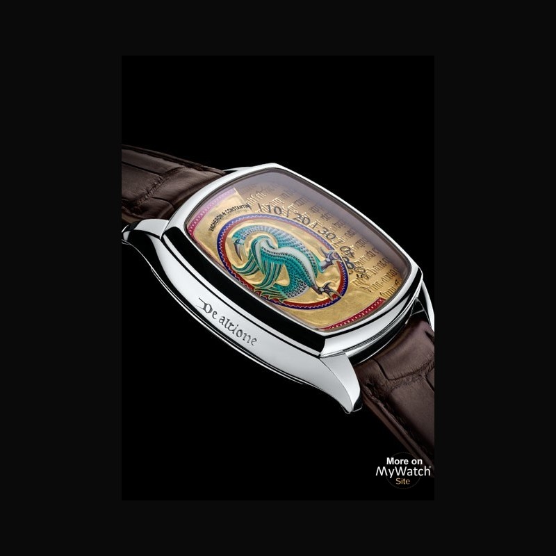
Watch Vacheron Constantin Métiers d'Art Savoirs Enluminés - Altion

Guy/uri_nlp_ner_workshop - 175bbd99d7dcc833049429d04334e7afe5763340
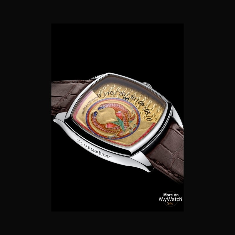
Watch Vacheron Constantin Métiers d'Art Savoirs Enluminés - Vultures
Light microscope (a – c and e) and μ -SXRF (d and f) images of

AVES

PDF) Retrograde Lymphatic Spread of Esophageal Cancer: A Case Report

AVES
Recommended for you
-
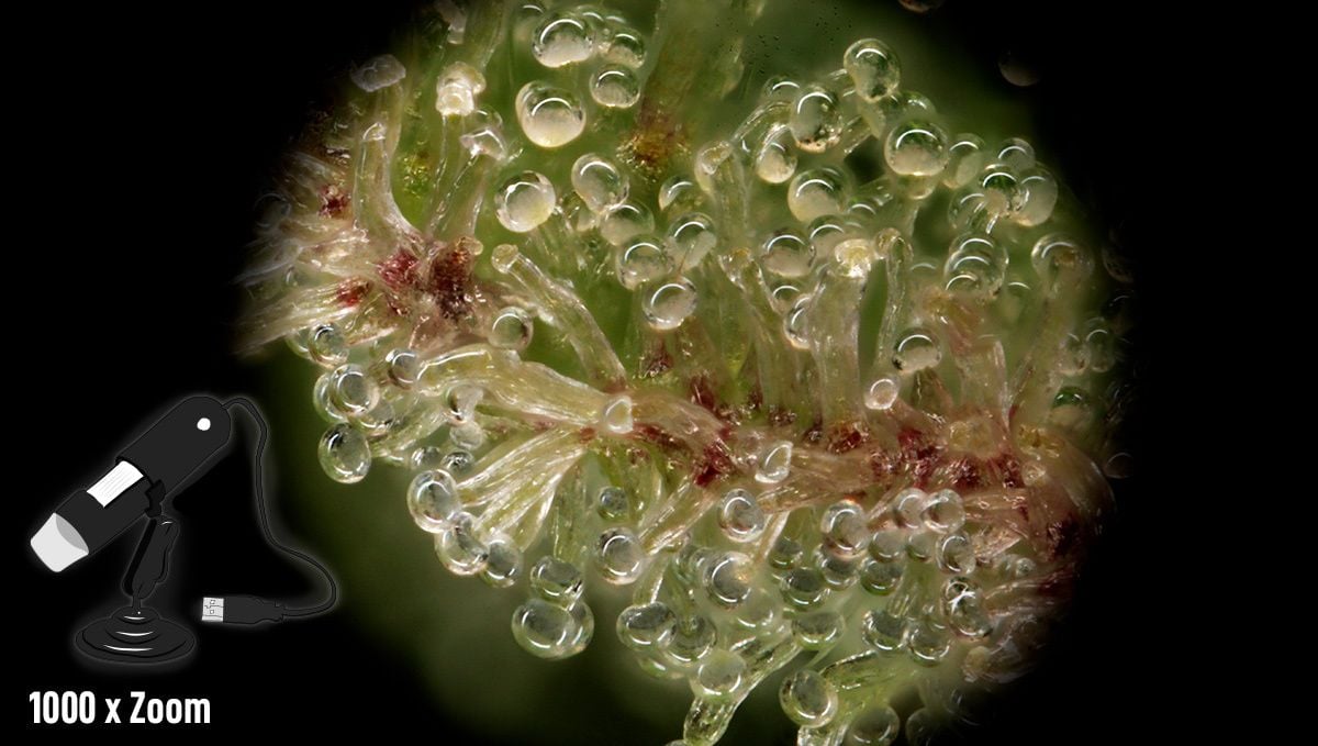 The Best Magnifiers to Identify Trichome Stages16 Jul 2024
 -
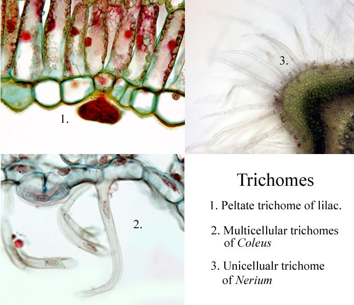 Three Types of Trichomes - UWDC - UW-Madison Libraries16 Jul 2024
 -
 Cannabis trichomes: what are they?16 Jul 2024
 -
![TOMLOV DM1S Wireless Digital Microscope [Easy and Fun] 50X-1000X 1080P HD WiFi Portable Handheld USB Trichome Mini Coin Microscope Camera Magnifier with Stand for iPhone iPad Android Phone & PC](https://m.media-amazon.com/images/I/71W0hsd4JZL.jpg) TOMLOV DM1S Wireless Digital Microscope [Easy and Fun] 50X-1000X 1080P HD WiFi Portable Handheld USB Trichome Mini Coin Microscope Camera Magnifier with Stand for iPhone iPad Android Phone & PC16 Jul 2024
TOMLOV DM1S Wireless Digital Microscope [Easy and Fun] 50X-1000X 1080P HD WiFi Portable Handheld USB Trichome Mini Coin Microscope Camera Magnifier with Stand for iPhone iPad Android Phone & PC16 Jul 2024 -
 SmartPhone Trichome Scope - Easiest way to check on Trichome Maturity16 Jul 2024
 -
 CUPAC ANATOMY SLIDE IMAGES16 Jul 2024
 -
 Trichomes de cannabis au microscope électronique- Alchimia Grow Shop16 Jul 2024
 -
 What is the absolute best trichome microscope out there16 Jul 2024
 -
GrowWeedEasy.com - I use a USB microscope to check trichomes16 Jul 2024
-
 Plant Leaf Trichome (hibiscus Sp.) Photograph by Dennis Kunkel16 Jul 2024

You may also like
-
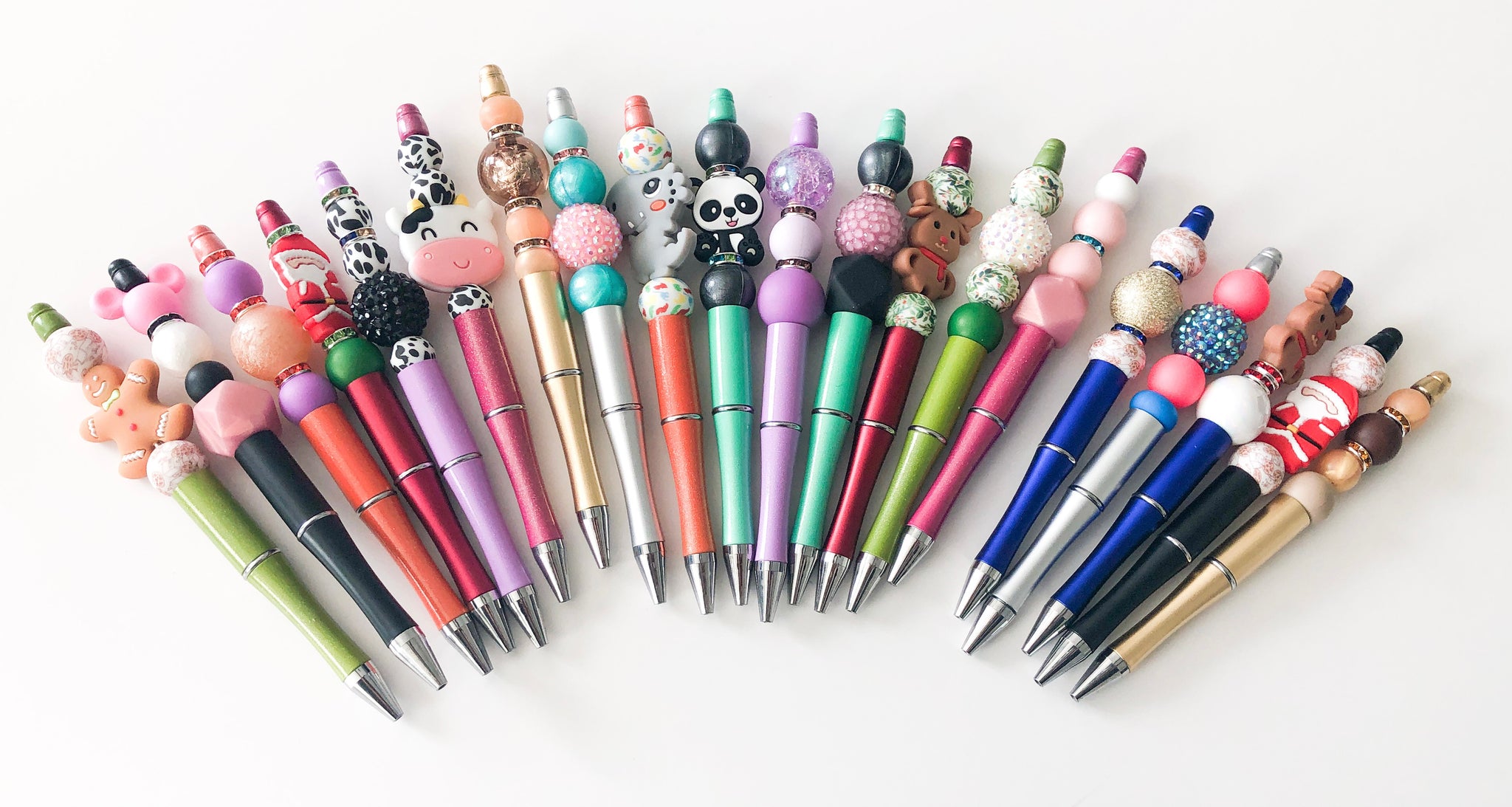 Bulk Order Beaded Pens16 Jul 2024
 -
 Official heights displayed at the Requiem : r/homestuck16 Jul 2024
 -
 Leather Dye Repair Kits - Auto Leather Repair Kits – Auto Leather Dye16 Jul 2024
 -
 Gunmtal O-rings Metal O Ring Spring Ring Clasp Push Gate O16 Jul 2024
 -
 Cute pencil cases: Cool pens storage to carry them in style + get organized16 Jul 2024
 -
 New 50Pcs White Wedding Car Decoration Gift Wrap Ribbon Bows Party Cars Chairs Ribbons Bows Delicate Kit16 Jul 2024
 -
 ebin lace bond spray vs glue|TikTok Search16 Jul 2024
 -
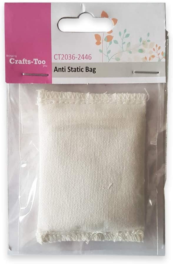 Crafts Too Anti-static Pouch for Heat Embossing / Card Making / Scrapbooking / Applying Glitter / Hand Lettering16 Jul 2024
 -
 French Fries Large - Order Online!16 Jul 2024
 -
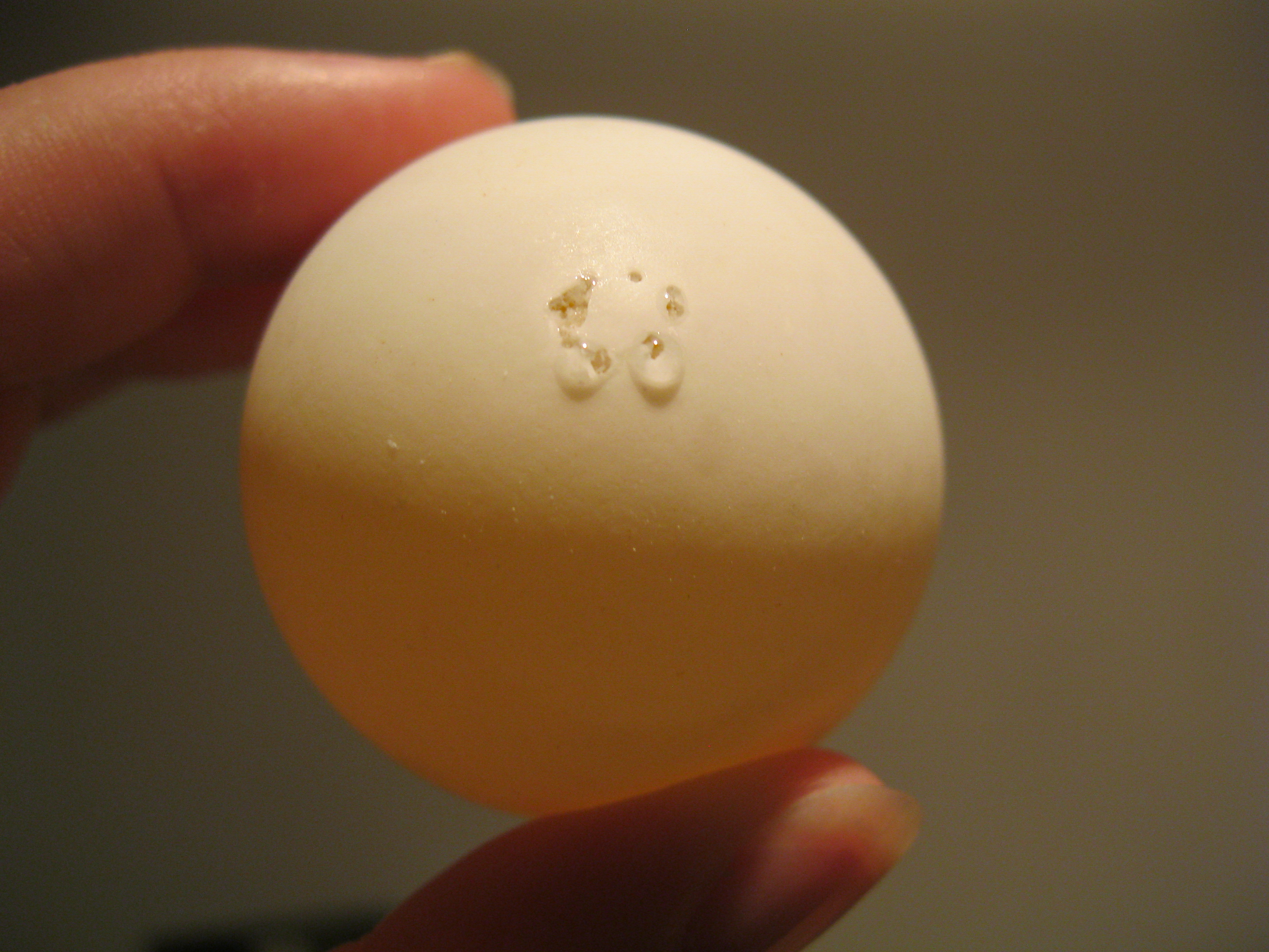 hollow eggs Change Here16 Jul 2024