Rose Petal Upper Surface, SEM - Stock Image - F017/4073 - Science Photo Library
By A Mystery Man Writer
Last updated 17 Jul 2024

Papillae on the upper surface of a rose flower petal (Rosa sp), coloured scanning electron micrograph (SEM). Papillae are projections from epidermal cells and in the rose they are conical in shape. DENNIS KUNKEL MICROSCOPY/SCIENCE PHOTO LIBRARY

Petal upper surface hi-res stock photography and images - Alamy
Rose Petal Upper Surface, SEM - Stock Image - F017/4073 - Science Photo Library

Rose petal surface. Each surface cell is 20 microns across! via @wellcomeimages

Focused ion beam scanning electron microscopic image of the rose petal

Rose Petal Upper Surface, SEM - Stock Image - F017/4073 - Science

Rose Petal Upper Surface, SEM - Stock Image - F017/4073 - Science

Rose Petal Upper Surface, SEM - Stock Image - F017/4072 - Science Photo Library

Focused ion beam scanning electron microscopic image of the rose petal

Advances in flexible sensors for intelligent perception system enhanced by artificial intelligence - Niu - 2023 - InfoMat - Wiley Online Library

Petal upper surface hi-res stock photography and images - Alamy

Rose Petal Iphone wallpaper images, Rose petals, Things under a microscope
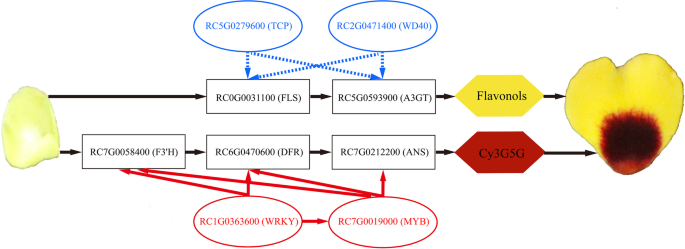
Metabolic profile and transcriptome reveal the mystery of petal blotch formation in rose, BMC Plant Biology

Rose Petal Lower Surface, SEM - Stock Image - C037/0543 - Science Photo Library

SEM image of the surface of the rose petal, revealing a periodic array

Focused ion beam scanning electron microscopic image of the rose petal
Recommended for you
-
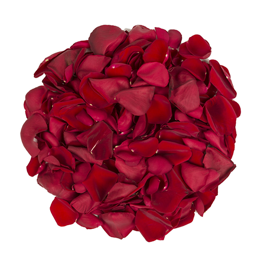
5,000 Rose Petals - Red17 Jul 2024
-
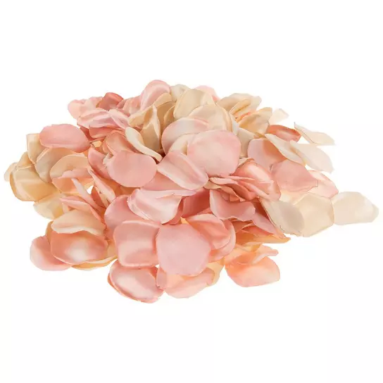
 GlobalRose 5000 Fresh Pink Rose Petals - Real Petals with Fast Delivery - Perfect for Valentine´s Day17 Jul 2024
 -
:max_bytes(150000):strip_icc()/rosepetaloil_recirc-c7e295acc9924e5da10a4a5f6b4ffd45.jpg) Rose Petal Oil Is an Under-the-Radar Ingredient With Benefits You Need to Know17 Jul 2024
Rose Petal Oil Is an Under-the-Radar Ingredient With Benefits You Need to Know17 Jul 2024 -
 rose petals Nutrition Facts and Calories, Description17 Jul 2024
 -
:max_bytes(150000):strip_icc()/9298909-e52c88030e3a44a1a340eaa7ea8fd9a3.jpg) Rose Petal Jam Recipe17 Jul 2024
Rose Petal Jam Recipe17 Jul 2024 -
Satin Rose Petal Filler17 Jul 2024
-
 2,212 Rose Petal Spread Images, Stock Photos, 3D objects, & Vectors17 Jul 2024
 -
 6,973 Rose Petal Stock Photos, High-Res Pictures, and Images - Getty Images17 Jul 2024
 -
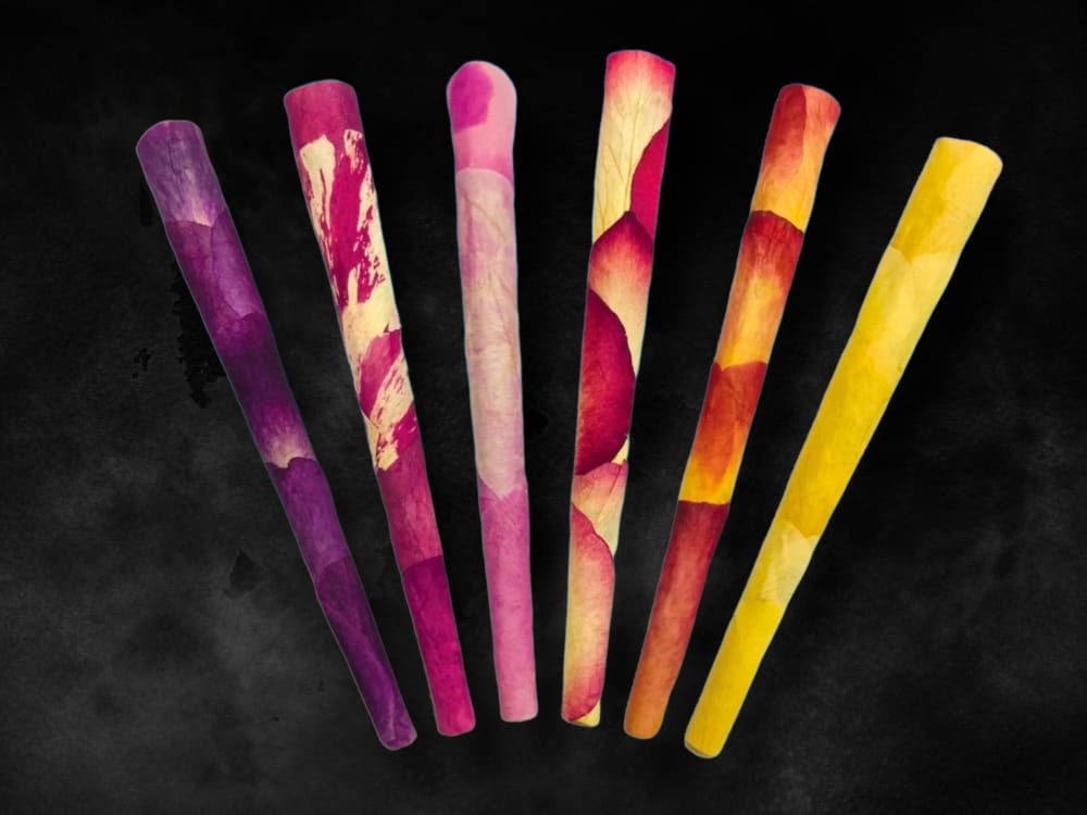 Best Rose Petal King Cones - 6 Pack17 Jul 2024
 -
 Rose Petal recipe ingredients - How to make a Rose Petal cocktail17 Jul 2024
You may also like
-
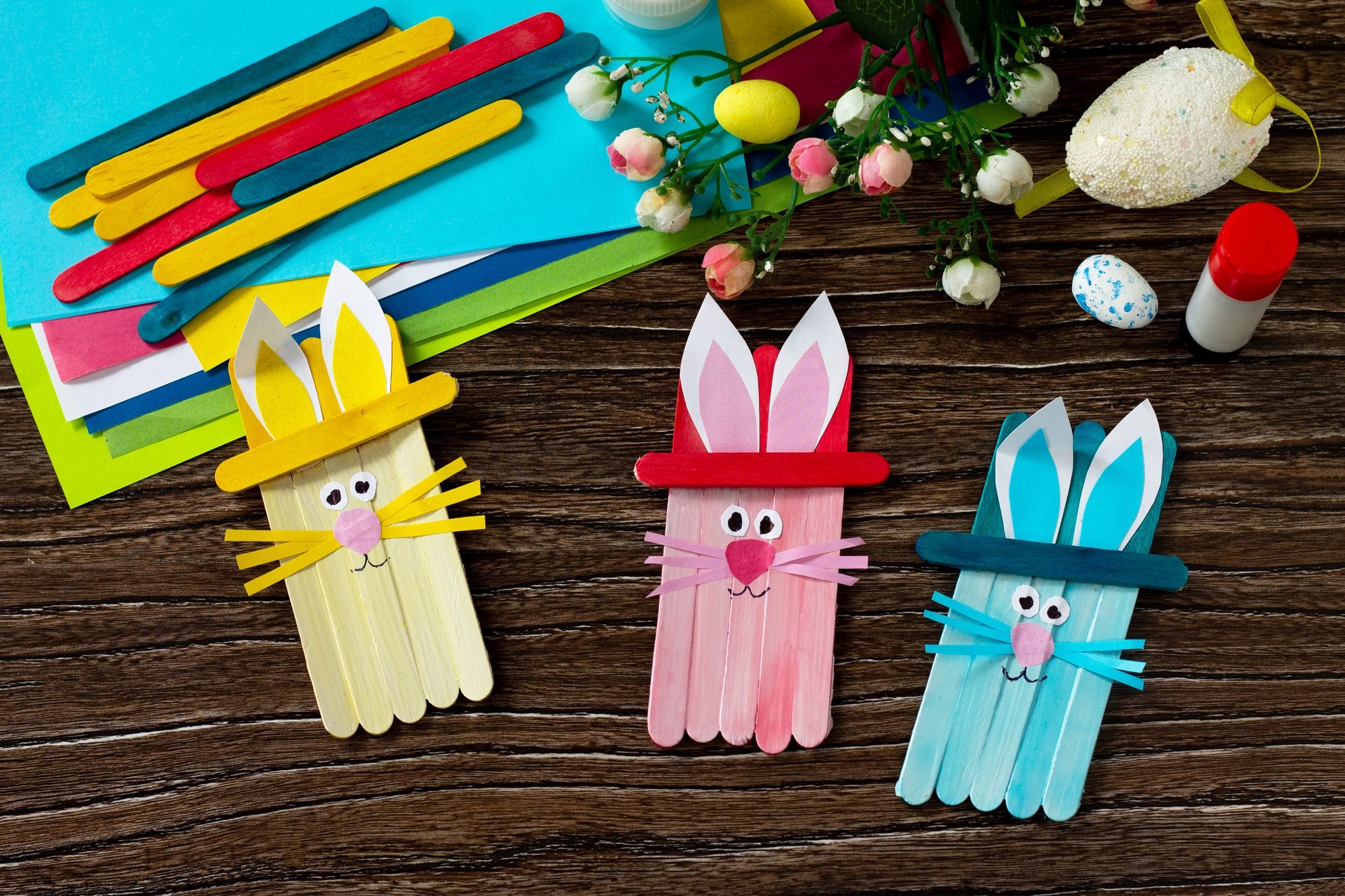 18 Easy Easter Crafts for Kids You Can Make At Home17 Jul 2024
 -
 FORTIVO Black Leather and Vinyl Repair Kit and Brown17 Jul 2024
 -
 1Pcs Long Dress Zipper Helper Puller Women Necklace Zipping Up Down Dress Boot Yourself Beaded Zipper Assistant Aid Tool17 Jul 2024
 -
 Sand Play for Child Development and Learning17 Jul 2024
 -
 NATURAL BAMBOO FOUNDATION BRUSH17 Jul 2024
 -
 Non Tarnish Gold Color Copper Craft Wire Assortment 16 to 24 Gauge17 Jul 2024
 -
 Wizard White Wax17 Jul 2024
 -
 Green Glitter Digital Background Texture — drypdesigns17 Jul 2024
 -
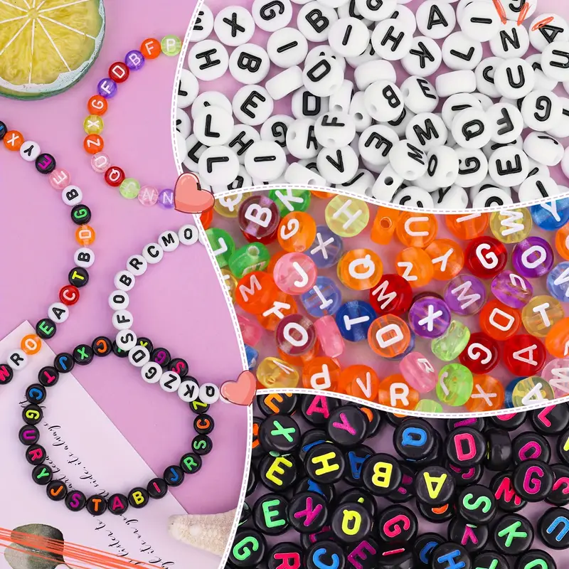 Girl Bracelet Making Set: Create Unique Jewelry Gifts For - Temu Philippines17 Jul 2024
 -
 Akoada Cute Canvas Roll Up Pencil Case with Pencil Pouch, Light Brown / Baby Crocodile17 Jul 2024