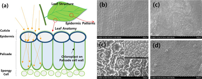
Scanning Eletron Microscopy photograph of the leaf surface of Solanum
By A Mystery Man Writer
Last updated 17 Jul 2024

Download scientific diagram | Scanning Eletron Microscopy photograph of the leaf surface of Solanum granuloso-leprosum Dunal. A – Unicelular tector trichomes; B – tector trichome, note that there are projections at the trichome base; C – tector trichome, note that there is a larger projection/ramification at the trichome base; D – tector trichome, note that there are two larger projection/ramification at the trichome base; E – tector trichome, note that there are three larger projection/ramification at the trichome base; F – tector trichome, note that there are four larger projection/ramification at the trichome base; G – tector trichome, note that there are five larger projection/ramification at the trichome base; H – tector trichome, note that there are six larger projection/ramification at the trichome base; I – tector trichome, note that there are eight larger projection/ramification at the trichome base; J – another angle from the six ramification tector trichome; and K – multicelular and multisseriated tector trichome, note the thick secondary cell wall. Scale Bars = 20 μm. from publication: Anatomy, histochemistry and micromorphology of leaves of Solanum granuloso-leprosum Dunal | In the present work the anatomical, histochemical and micromorphological features of S. granuloso-leprosum leaves were approached in order to evaluate its characteristics associated with its pioneer role. Glandular and non-glandular trichomes were observed on both epidermal | Micromorphology, Solanum and Plant Anatomy | ResearchGate, the professional network for scientists.
Omnidirectional Light Capture by Solar Cells Mimicking the Structures of the Epidermal cells of Leaves

SEM micrographs showing surface structure of chemical fixed leaves and
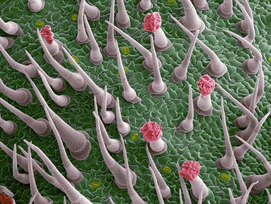
Tomato Leaf Surface, Sem Photograph by Steve Gschmeissner - Pixels

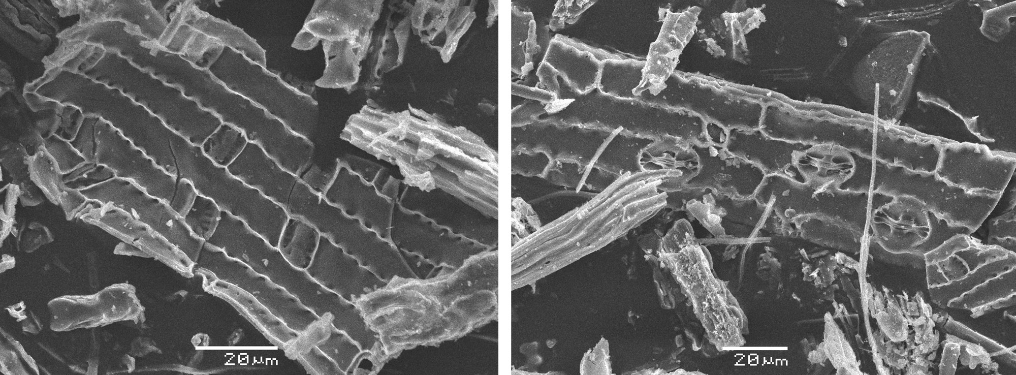
Scanning electron micrographs of the leaf of Solanum elaeagnifolium.

Morphological characterisation of trichomes

Scanning electron microscopic images of the lower leaf epidermis

Herbs Micronaut: The fine art of microscopy by science photographer Martin Oeggerli
Types of evidence used to study ancient grasses - Earth@Home: Evolution

PDF] Plant trichomes and the biomechanics of defense in various systems, with Solanaceae as a model
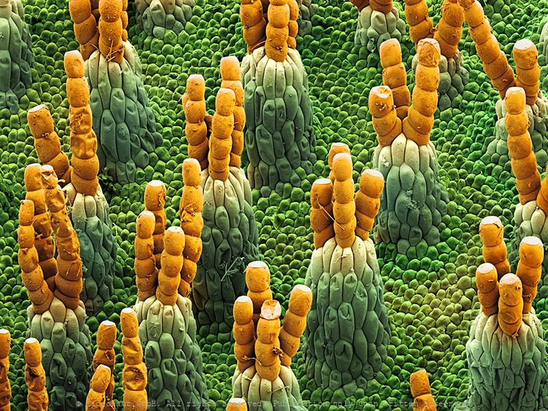
Herbs Micronaut: The fine art of microscopy by science photographer Martin Oeggerli

Plants, Free Full-Text

Scanning electron micrographs of the leaf of Solanum elaeagnifolium.
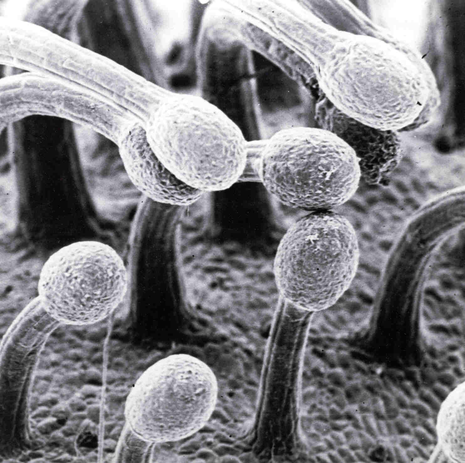
Image library - substance type - Quorum Technologies Ltd
Scanning electron microscopic (SEM) images captured at 60×
Recommended for you
-
 Sap secretion in late flower, looks like an oddly shaped trichome. : r/ microscopy17 Jul 2024
 -
 What is the absolute best trichome microscope out there? - Harvesting - I Love Growing Marijuana Forum17 Jul 2024
 -
 Cannabis Trichomes Overview17 Jul 2024
 -
 Scanning electron microscope micrographs of trichomes of tribe17 Jul 2024
 -
![TOMLOV DM1S Wireless Digital Microscope [Easy and Fun] 50X-1000X 1080P HD WiFi Portable Handheld USB Trichome Mini Coin Microscope Camera Magnifier](https://m.media-amazon.com/images/I/71W0hsd4JZL.jpg) TOMLOV DM1S Wireless Digital Microscope [Easy and Fun] 50X-1000X 1080P HD WiFi Portable Handheld USB Trichome Mini Coin Microscope Camera Magnifier17 Jul 2024
TOMLOV DM1S Wireless Digital Microscope [Easy and Fun] 50X-1000X 1080P HD WiFi Portable Handheld USB Trichome Mini Coin Microscope Camera Magnifier17 Jul 2024 -
 Trichome microscope hack for crystal clear pics with 100% stillness! - Harvesting - I Love Growing Marijuana Forum17 Jul 2024
 -
 Best Microscope For Viewing Trichomes: Top 317 Jul 2024
 -
 Plant Leaf Trichome (hibiscus Sp.) Photograph by Dennis Kunkel Microscopy/science Photo Library - Pixels17 Jul 2024
 -
 Trichome on leaf of Arabidopsis thaliana17 Jul 2024
 -
 File:Müürlooga (Arabidopsis thaliana) lehekarv (trihhoom) 311 080417 Jul 2024
You may also like
-
 5D Diamond Painting Marvel Super Hero Collection Kit - Bonanza17 Jul 2024
 -
 Best Painter's Tape: How to Choose the Best Type for Your Project17 Jul 2024
 -
 Post-It® Easel Pad Plain, 25 X 30 (Pack of 4)17 Jul 2024
 -
 Rub'N'Etch Stencils - Armour Products.com - Wholesale Glass Etching Supplies17 Jul 2024
 -
Simmi chain mail halterneck plunge front side silt mini dress in gold17 Jul 2024
-
 Arcilla para modelar 1,5 kg la unidad17 Jul 2024
 -
 Printable Shrink Plastic Jewelry Making, Shrinking InkJet Plastic Fil, MiniatureSweet, Kawaii Resin Crafts, Decoden Cabochons Supplies17 Jul 2024
 -
 Wooden Craft Board Lap Desk Board Painting Board17 Jul 2024
 -
 Unlock your Potential: JUMBO Custom Diamond Painting Kit - Full17 Jul 2024
 -
 Gerich Car Bling Steering Wheel Cover for Women Girls Burgundy 15 inch Universal Red Crystal Rhinestone Diamonds Bling Bling Accessories Anti-Slip17 Jul 2024